


Custom PNA Oligos
Custom PNA oligomers can be unlabeled or labeled by dyes or other chemicals. They can also be conjugated to peptides to add other functionality. Linkers can be added as a spacer and also to improve solubility. Gamma PNA or modified bases can increase Tm and specificity of PNA even further. All our PNA products are provided >95% purity, accompanied by a COA including HPLC purity and mass data.
- Technology
- PNA Applications
- PNA Design Guidelines
- PNA Handling Guidelines
- Unlabeled PNA
- Labeled PNA
- PNA-Peptide Conjugates
- Gamma PNA
- Publicatons
- Order
About PNA
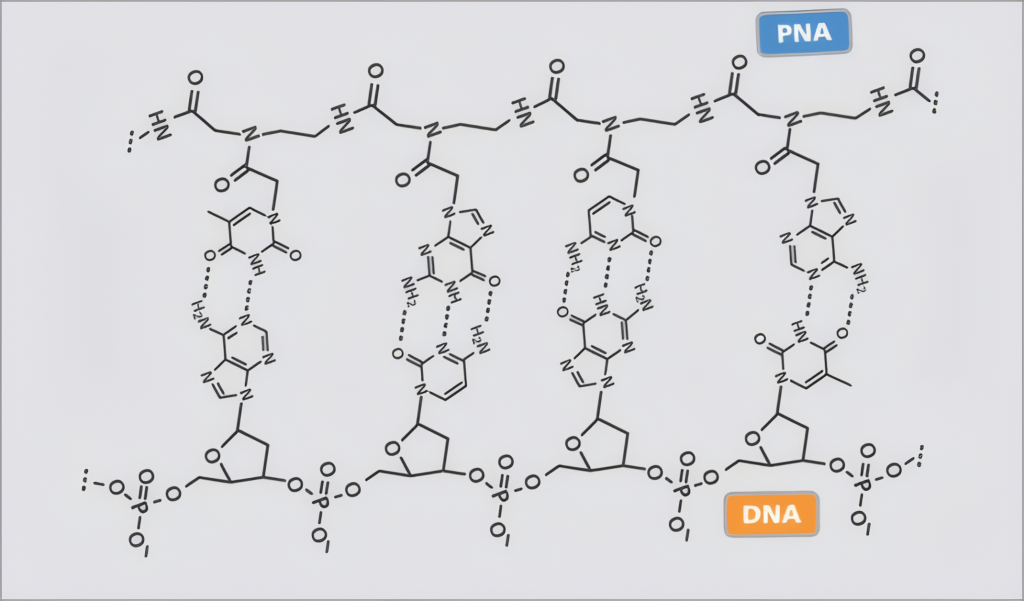
PNA (Peptide Nucleic Acid) is a synthetic polymer that mimics the structure of DNA and RNA. In PNA, the nucleobases are attached to the backbone by methylene carbonyl bonds as in peptides. Unlike DNA or RNA, PNA lacks negatively charged phosphate groups in its backbone. This absence of charge results in stronger binding between PNA and DNA or PNA and RNA, compared to DNA-DNA or DNA-RNA interactions, as there is no electrostatic repulsion to overcome. PNA molecules exhibit remarkable chemical stability both in vitro and in vivo. They are also resistant to degradation by DNases, RNases, and proteases.
PNA Advantages
- High affinity, fast binding due to neutral backbone
- High specificity (SNP detection)
- Salt independent binding
- Chemical stability at high temperature and various pH
- Resistant to RNases, DNases, and proteases
Due to its neutral backbone, PNA binds to DNA and RNA with higher affinity and specificity. The melting temperature (Tm) of a PNA/DNA duplex is typically about 1°C higher per base pair compared to a DNA/DNA duplex. Because of this strong binding, PNA oligomers are effective even at shorter lengths, with most commonly used sequences ranging from 13 to 20 bases. PNA/RNA duplexes exhibit an even higher Tm, exceeding that of PNA/DNA duplexes by a few degrees.
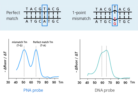
PNA/DNA hybrids exhibit a greater binding affinity difference between perfect matches and mismatches than DNA/DNA hybrids. For example, a single-base mismatch typically lowers the melting temperature (Tm) by about 15°C in a PNA/DNA hybrid, compared to about 10°C in a DNA/DNA hybrid. This enhanced sensitivity makes PNA probes highly effective for detecting even single nucleotide mismatches.
For more guidelines and Tm calculations, please refer to PNA TOOL.
For comprehensive information, the PNA oligomer design guidelines are available for download. 
Due to high affinity and specificity, PNA has superior hybridization efficiency and sensitivity.
PNA Applications
- Sequence specific PCR blocker (PNA clamp)
- FISH probes for telomere, centromere, gene specific probes, microorganisms
- Anti-sense/ anti-microbial reagents
- miRNA inhibitors
- Double strand DNA invasion & capture
- Microarray probes
- Quantification of RNA therapeutics
- Antisense and antigene therapy
PNA can bind with high specificity to complementary RNA or DNA sequences, blocking translation or transcription. This makes it a promising candidate for gene silencing or correction in therapeutic applications.
- siRNA quantification
PNA probes can be used in hybridization-based assays (e.g., LC-FD) for highly specific detection and quantification of siRNA in biological samples.
- PCR clamping and blocking
PNAs can suppress amplification of unwanted sequences (e.g., wild-type alleles) during PCR, allowing selective amplification of rare or mutant alleles with high sensitivity.
- FISH (Fluorescence In Situ Hybridization)
PNA probes are used in FISH for detecting specific DNA sequences in chromosomes with high signal-to-noise ratio and rapid hybridization, making them ideal for telomere or centromere analysis.
- PNA–peptide conjugates
Linking PNA to cell-penetrating peptides (CPPs) facilitates intracellular delivery for gene targeting or antimicrobial applications. Conjugates can also include nuclear localization signals or other functional moieties.
- Double-stranded DNA invasion & capture
PNA can invade double-stranded DNA by forming stable PNA–DNA–PNA triplexes or duplexes, displacing one DNA strand. This allows highly specific binding without prior denaturation and is useful for gene targeting, mutation detection, or isolating genomic regions for downstream analysis.
- PNA molecular beacons
These are PNA probes labeled with a fluorophore and quencher that fluoresce upon hybridization, enabling real-time detection of specific nucleic acid targets in live cells or diagnostics.
- Molecular diagnostics
Due to its strong binding and mismatch discrimination, PNA is widely used in detecting single nucleotide polymorphisms (SNPs), mutations, or pathogen sequences in clinical diagnostics.
- Biosensors and nanotechnology
PNA’s stability and specificity make it an excellent component for biosensors, often immobilized on surfaces or nanoparticles to detect target nucleic acids in complex samples.
For aqueous-based applications such as PCR, it is best to use sequences with purine content under 50%, G content below 35%, and total length under 23 bases. For applications involving organic solvents such as FISH or FD-LC, purine content should be 75% or lower. If your target is double-stranded DNA, consider using the reverse complement sequence.
Linkers are commonly used as a spacer when conjugating PNA with other functional groups like peptides or dyes. Hydrophilic linkers such as O, E, and X linkers can enhance the solubility of PNA oligomers. Several types of linkers are possible. The O linker (also known as AEEA or eg1) is most commonly used spacer, which also improve solubility. Other spacers such as E, X, C3, C4, C6, and C12 linkers are also available. C6SH or C11SH can be used for adding a thiol group to the PNA.
If you have no flexibility in PNA sequence, adding two lysines or O-linkers can improve solubility.
Possible linkers
- O linker (also called AEEA or eg1): improve solubility, most commonly used
- As a spacer: O, E, X, L35, C4, C6, C12 linker
- Thiol addition: C6SH, C11SH
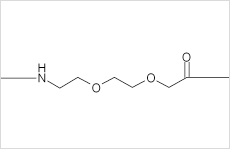 O-linker (9 atoms, 1.3 nm) |
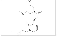 E-linker |
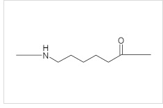 C6A linker (7 atoms, 1.1 nm) |
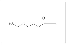 C6SH |
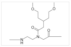 X-linker |
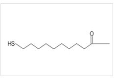 C11SH (12 atoms, 1.9 nm) |
To calculate Tm and check other properties, please use PNA Tool.
If you are selecting a PNA oligomer from a target region longer than 20 bases, PNA Designer Tool can help narrow down optimal sequences.
Our team is happy to review your chosen sequences or assist with custom design. Feel free to contact us at order@pnabio.com.
General Guidelines for PNA Oligomers
- Design the PNA oligomer to be complementary to the intended DNA or RNA target, just like standard DNA primers. For double-stranded DNA targets, either strand can be used. When possible, evaluate both orientations and select the sequence with the most favorable binding and solubility characteristics.
- PNA oligomers bind to complementary DNA or RNA targets with significantly higher affinity than standard oligonucleotides. The Tm of PNA is about 1°C per base pair higher than DNA. There is about a 15°C Tm difference for a single-base mismatch for PNA, while it is about 10°C for DNA. PNA binds most strongly to a complementary PNA target, followed by RNA and then DNA.
- PNA with a high purine content (>50%), a high amount of G bases (>35%), a long purine stretch (>7 bp), or a longer length (>30 bp) may exhibit reduced solubility in aqueous solutions. For applications limited to aqueous solution, keep these constraints in mind. For applications utilizing organic solvents, such as FISH, these constraints do not apply.
Solubility Tip
Consider adding solubility enhancers such as two lysine residues or O linkers. For double-stranded DNA targets, reverse-complement sequences can be used to avoid high purine content.
- Due to the neutral backbone of PNA, the binding of PNA to nucleic acid is less dependent on ionic strength and pH. PNA binding to DNA or RNA is not affected significantly from 10 mM to 1 M NaCl concentration.
- Please refer to the PNA Tool on our website for design guidelines and Tm calculation.
Handling and Storage of PNA Oligomers Upon Receipt
- Briefly centrifuge the tube before opening.
- Dissolve PNA to prepare a 20–100 µM stock. For example, dissolving 50 nmol of PNA in 500 µL of solvent results in a 100 µM stock. Water is generally the recommended solvent for PNA. For FISH, formamide is a better alternative. Avoid Tris buffer, as PNA tends to have lower solubility.
- To help ensure complete dissolution, heat the PNA solution at 55°C for 5 minutes and mix thoroughly. If the PNA is not fully dissolved, heat the PNA at 85°C for an additional 5 minutes. Lowering the PNA concentration may also improve solubility. At typical working concentrations of 1 µM or lower, most PNAs should remain soluble in aqueous assay solutions.
-
If the PNA is still not fully dissolved, consider the following options:
- For applications that can include organic solvents, add DMSO or DMF to a final concentration of 10–20%, or up to 50%. Repeat the heating step if necessary.
- For applications that cannot use organic solvent, add glacial acetic acid to a final concentration of 0.1–1% v/v.
- For the most difficult PNA to dissolve, 0.1% trifluoroacetic acid, TFA, v/v and 20% DMSO may be used.
- Store in aliquots at 4°C for short-term use, for up to one month. Storing PNA at −20°C or −70°C is recommended for long-term storage. Protect fluorescently labeled probes from light.
- After taking the PNA solution out of cold storage, heat it at 55°C for 5 minutes and mix thoroughly before use.
- PNA is highly stable and should remain intact for more than three years when properly stored.
Recommended Stock Preparation
- Recommended stock concentration: 20–100 µM
- Recommended solvent: water
- For FISH applications: formamide may be used
- Avoid Tris buffer when possible
- Typical working concentration: 1 µM or lower
PNA Quantification
Calculate the molar extinction coefficient, ε260, of the PNA oligomer. If the PNA is conjugated to a fluorophore or another chemical that contributes to absorbance at 260 nm, include its contribution in the calculation.
Extinction Coefficient Formula
ε260 (PNA oligomer) = Σ εi × ni
- i = A, G, C, and T
- εi = molar extinction coefficient of the base
- ni = number of each base
| Base | ε260 [mL/(µmol × cm)] |
|---|---|
| A | 13.7 |
| C | 6.6 |
| G | 11.7 |
| T | 8.8 |
- Calculate the molar extinction coefficient, ε260, of the PNA oligomer. Include the contribution of a fluorophore or other chemical modification if it contributes to absorbance at 260 nm.
- If needed, transfer a known volume of the PNA stock solution and dilute it to an appropriate final volume. Calculate the dilution factor.
- Measure the absorbance of the PNA solution at 260 nm using a spectrophotometer.
- Calculate the PNA concentration using the formula below.
PNA Concentration Formula
PNA concentration (mM) = [A260 ÷ (ε260 × path length in cm)] × dilution factor
Due to its high specificity and affinity, PNA oligomers effectively bind to their target nucleic acids. Unlabeled PNAs can be used as gene-specific blockers in PCR reactions (PNA clamping). PNA clamping can efficiently detect SNP mutations in a target gene as PNA can distinguish a single base mismatch.
When designing PNA oligomers for use in aqueous solution-based applications such as PCR, the following guidelines are recommended to ensure good solubility:
- Purine content <50%
- G bases <35%
- PNA length <23 bp
- Purine stretches <6 residues
For double-strand DNA targets, a reverse complement sequence can be chosen for PNA design. If there is no flexibility in PNA design, solubility enhancers such as two O linkers or two Lysines can be incorporated into the PNA.
Please refer to PNA TOOL for Tm predictions and other guidelines.
To learn more about PNA clamping technology and the PNA clamp products available as catalog items, please visit PCR blockers.
Our technical team would be happy to review your PNA sequences or help with the PNA design. Quotation requests, inquiries, and orders can be directed to order@pnabio.com.
A wide range of labels are available, including fluorophores such as FAM, FITC, Alexa dyes, Atto dyes, Cy dyes,HEX, ROX, Thiazole Orange (-NHS ester or carboxy amide), as well as quenchers like BHQ and Dabcyl, and other functionalities such as Acridine, Alkyne (DBCO or Pentynoic acid), Azide, Biotin, Digoxigenin, Ferrocene, Maleimide, Methylene Blue, Myristol, Palmitic acid, Pacific Blue, Thiol, and more.
A PNA probe with a fluorophore at the 5′ end and a quencher at the 3′ end can function as a molecular beacon (MB). Unlike DNA, the flexible structure of PNA allows it to remain in a quenched state without requiring a stem-loop design in unbound form, making probe design shorter and simpler. A wide range of fluorophore and quencher pairs are available. Thanks to the high specificity and binding affinity of PNA, molecular beacons offer highly sensitive detection without the need to wash away unbound probes.
For custom conjugation, please send an email to order@pnabio.com.
| Dye | Ex (nm) | Em (nm) | Dye | Ex (nm) | Em (nm) |
| FAM | 492 | 518 | Atto425 | 436 | 484 |
| FITC | 492 | 518 | Alexa Fluor 488 | 494 | 517 |
| Oregon Green 488 | 498 | 526 | TAMRA | 553 | 576 |
| Cy3 | 550 | 570 | Texas Red | 596 | 615 |
| Alexa Fluor 546 | 556 | 573 | Atto550 | 554 | 574 |
| Alexa Fluor 532 | 530 | 555 | Atto647N | 644 | 667 |
| Cy5 | 650 | 670 | Alexa Fluor 647 | 650 | 668 |
| Thiazol Orange | 510 | 530 | Dabcyl | 454 | |
| JOE | 520 | 548 | BHQ-2 | 560-670 | |
| BHQ-1 | 480-580 | BHQ-3 | 620-730 |
Because the PNA backbone is polyamide-based, it can be readily conjugated to peptides for added functionality. For instance, adding lysine residues can improve solubility, while cysteine can enable conjugation via disulfide bond formation.
Peptides may be attached to either the 5′ or 3′ end of the PNA, with an O-linker optionally included as a spacer between the peptide and the PNA strand.
PNA–peptide conjugates have a range of applications. In antimicrobial research, PNA sequences (typically 10–15 bases) targeting essential microbial genes can be paired with cell-penetrating peptides (CPPs) like (KFF)3K or (RXR)4XB for intracellular delivery. In mammalian cells, antisense PNAs are often designed to target mRNA regions near the 5′ ATG start codon. In addition, nuclease localization signl (NLS) can be conjugated to facsilitate transport of PNA into nucleus. Bipartite PNA-peptide can also efficiently capture target microorganisms.
We can synthesize PNA-peptide conjugates up to 50 residues in length via standard linear synthesis. For longer constructs, PNA and peptide can be synthesized separately and conjugated by maleimide-cysteine ligation.
For inquiries or to place an order, please contact us at order@pnabio.com.
Gamma PNA contains an R group at a stereogenic center at the γ-carbon of the N-aminoethyl glycine unit. The addition of gamma modifications increases the melting temperatures (Tm) by 5-8 °C , enhancing sequence-specific binding and enabling invasion into double-stranded DNA. Gamma PNA also provides benefits such as improved solubility, reduced self-aggregation, stable duplex formation with DNA, and flexibility for multi-labeling and other functionalization.
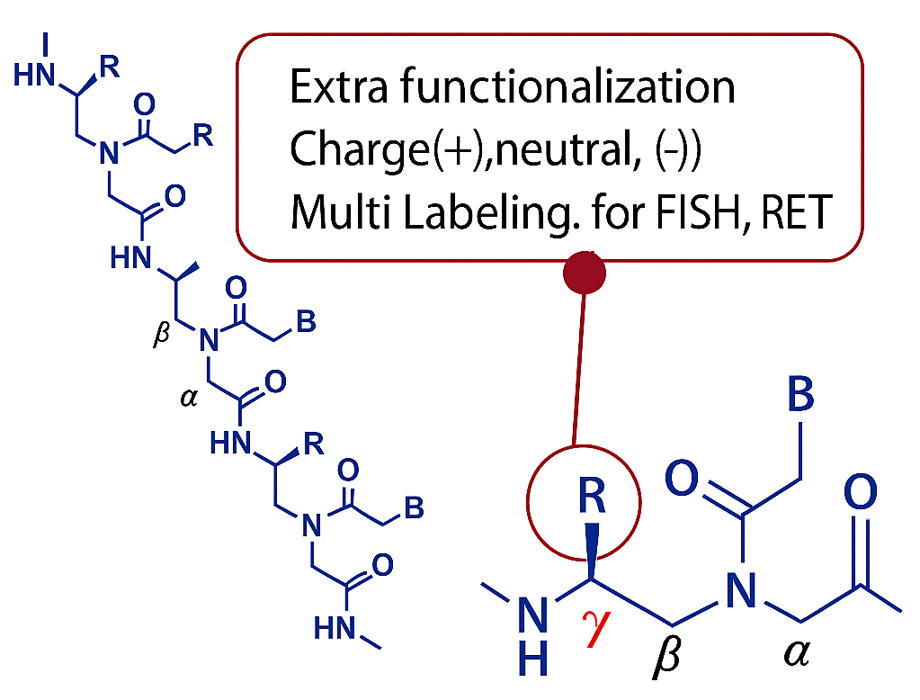
- Possible gamma functional groups
– Lysine: better solubility, possible for dual-labeling, the potential for cell penetration
– Alanine and glutamic acid are also possible modifications.
– Lysine: enhance solubility, possible for dual-labeling, the potential for cell penetration
– Glutamic acid: adds negative charges, improves solubility
– Alanine: increases hydrophobicity, can be added to every residue
– Serine and miniPEG: improves solubility without altering charges
Sensitive siRNA quantification
- Evaluating the effects of aging on distribution and gene silencing activity of lipid-siRNA conjugates delivered into cerebrospinal fluid. Ligocki AP et al. (2026) Mol Ther 34(9):5366–5381.
- Intradermal delivery of lipophilic siRNAs enables prolonged skin retention and sustained gene silencing in a porcine model. Fakih HH et al. (2026) Nat Commun 17:2292.
- Intravenous lipid-siRNA conjugate mediates gene silencing at the blood-brain barrier and blood-CSF barrier. Sorets AG et al. (2025) J Control Release 387:114226.
- Elucidating brain transport pathways and cell type-dependent gene silencing of a durable lipid–siRNA conjugate administered into cerebrospinal fluid. Sorets AG et al. (2025) Nucleic Acids Res 53(12):gkaf600.
- A programmable dual-targeting siRNA scaffold supports potent two-gene modulation in the central nervous system. Belgrad J et al. (2024) Nucleic Acids Res 52(11):6099–6113.
- Rational design of a JAK1-selective siRNA inhibitor for the modulation of autoimmunity in the skin. Tang Q et al. (2023) Nat Commun 14:7099.
- A novel hybridization LC-MS/MS methodology for quantification of siRNA in plasma, CSF and tissue samples. Yuan L et al. (2023) Molecules 28(4):1618.
- Using Peptide Nucleic Acid Hybridization Probes for Qualitative and Quantitative Analysis of Nucleic Acid Therapeutics by Capillary Electrophoresis. Hutanu A et al. (2023) Analytical Chemistry 95(11):4914–4922.
- Docosanoic acid conjugation to siRNA enables functional and safe delivery to skeletal and cardiac muscles. Biscans A et al. (2021) Molecular Therapy 29(4):1382–1394.
- Quantitative determination of a siRNA (AD00370) in rat plasma using peptide nucleic acid probe and HPLC with fluorescence detection. Tian Q et al. (2017) Bioanalysis 9(11):861–872.
Anti-sense PNA
- Targeting Oncogenic KRAS Using Peptide Nucleic Acid Oligomers Attached to Cell-Penetrating Peptides. Mondal J et al. (2026) Int J Mol Sci 27(16):7158.
- A secreted endosymbiont protein essential for colonizing host cells. Maeda GP et al. (2026) Nature 656:443–451.
- Programmable antisense oligomers for phage functional genomics. Gerovac M et al. (2025) Nature 646:1195–1203.
- Broad Host Range Peptide Nucleic Acids Prevent Gram-Negative Biofilms Implicated in Catheter-Associated Urinary Tract Infections. Karp HQ et al. (2025) Microorganisms 13(8):1948.
- Endosomolytic Peptides Enable the Cellular Delivery of Peptide Nucleic Acids. Giancola JB & Raines RT (2024) Chem Commun (Camb) 60(100):15019–15022.
- Antisense inhibition of RNA polymerase α subunit of Clostridioides difficile. Pal R & Seleem MN (2023) Microbiol Spectr 11(5):e0175523.
- Antimicrobial activity of antisense peptide–peptide nucleic acid conjugates against non-typeable Haemophilus influenzae in planktonic and biofilm forms. Otsuka T et al. (2017) J Antimicrob Chemother 72(1):137–144.
- Dual Repression of the Multidrug Efflux Pump CmeABC by CosR and CmeR in Campylobacter jejuni. Grinnage-Pulley T et al. (2016) Front Microbiol 7:1097.
PNA clamping
- Climate and temporal variation in bacterial and fungal communities of the rhizosphere and roots of young avocado trees. Zaman M et al. (2026) Biol Fertil Soils.
- Clearing the Noise: Seasonal Dynamics of Endophytic Bacteria in Fagus sylvatica Leaves Revealed by Application of PNA Clamps. Giubilei I et al. (2026) Physiol Plant 178:e70897.
- Peptide nucleic acid (PNA) clamps enhance root microbiome profiling in wheat and maize. Mukhtar H et al. (2025) Environ Microbiome 20:148.
- Nutritional and microbiological dynamics in the preparation of prahoc fish paste. Thanh C et al. (2025) PLoS One 20(4):e0321834.
- Influence of genotype, nodule position, and edaphic factors on microbial diversity and assembly of pigeonpea (Cajanus cajan) root nodules in Indian soils. Basu A et al. (2025) Environ Microbiome 20:41.
- Bacterial community structure analysis on Listeria monocytogenes inoculated spinach leaves is affected by PCR based methods to exclude chloroplast co-amplification. Culliney P & Schmalenberger A (2025) The Microbe 6:100258.
- Peptide nucleic acid (PNA) clamps reduce amplification of host chloroplast and mitochondria rRNA gene sequences and increase detected diversity in 16S rRNA gene profiling analysis of oak-associated microbiota. Hussain U et al. (2025) Environ Microbiome 20:14.
- Influence of Growth Support on the Diversity, Composition, and Functionality of Microbial Communities Associated with Tillandsia recurvata. Siqueira JS et al. (2024) Microb Ecol 87:129.
- Peptide Nucleic Acid-Mediated Regulation of CRISPR-Cas9 Specificity. Carufe KEW et al. (2024) Nucleic Acid Ther 34(5):245–256.
- Conservation and diversity of the pollen microbiome of Pan-American maize using PacBio and MiSeq. Khalaf EM et al. (2023) Front Microbiol 14:1276241.
- An amplification-free, 16S rRNA test for Neisseria gonorrhoeae in urine. Zheng Z et al. (2023) Sensors Diagn 2(1):163.
- Seagrass (Zostera marina) transplant experiment reveals core microbiota and resistance to environmental change. Adamczyk EM et al. (2022) Mol Ecol 31:5107–5123.
- Bacterial Communities Show Algal Host (Fucus spp.) Zone Differentiation Across the Stress Gradient of the Intertidal Zone. Quigley CTC et al. (2020) Front Microbiol 11:563118.
PNA biosensors
- Programmable PNA–nanoparticle hybrids as nanoscale recognition architectures for amplification-free nucleic acid recognition. Natarajan S et al. (2026) Biosens Bioelectron 297:118380.
- PNA Functionalized Gold Nanoparticles on TiO2 Nanotubes Biosensor for Electrochemical DNA Fragment Detection. Kim Y & Mohanty SK (2025) Adv Mater Interfaces 12(8):2400762.
- Isothermal biosensor for highly specific detection of single-base mutations based on peptide nucleic acid and graphene oxide. Park SM et al. (2025) Sens Actuators B Chem 440:137880.
- A highly sensitive and robust electrochemical biosensor for microRNA detection based on PNA-DNA hetero-three-way junction formation and target-recycling catalytic hairpin assembly amplification. Yu X et al. (2024) Talanta 266:125020.
- Magnetic Nanoparticle-Based Electrochemical Sensing Platform Using Ferrocene-Labelled Peptide Nucleic Acid for the Early Diagnosis of Colorectal Cancer. Hanoglu SB et al. (2022) Biosensors (Basel) 12(9):736.
- PNA microprobe for label-free detection of expanded trinucleotide repeats. Asefifeyzabadi N et al. (2022) RSC Adv 12(13):7757–7761.
- Detection of Tumor DNA in Human Plasma with a Functional PLL-Based Surface Layer and Plasmonic Biosensing. Bellassai N et al. (2021) ACS Sens 6(6):2307–2319.
- Paper-based electrochemical peptide nucleic acid (PNA) biosensor for detection of miRNA-492: a pancreatic ductal adenocarcinoma biomarker. Moccia M et al. (2020) Biosens Bioelectron 165:112371.
- E-DNA scaffold sensors and the reagentless, single-step, measurement of HIV-diagnostic antibodies in human serum. Parolo C et al. (2020) Microsyst Nanoeng 6:13.
- Fully Dried Two-Dimensional Paper Network for Enzymatically Enhanced Detection of Nucleic Acid Amplicons. Byers KM et al. (2020) ACS Omega 5(9):4673–4681.
- Multiplex Paper-Based Colorimetric DNA Sensor Using Pyrrolidinyl Peptide Nucleic Acid-Induced AgNPs Aggregation for Detecting MERS-CoV, MTB, and HPV Oligonucleotides. Teengam P et al. (2017) Anal Chem 89(10):5428–5435.
PNA FISH
- MUTYH Activity Maintains Telomere Stability in Response to Chronic Telomeric 8-Oxoguanine Damage in Cancer Cells. De Rosa M et al. (2026) Biomolecules 16(9):1234.
- Distinct ATRX functions cooperate with 9-1-1 and CST complexes to safeguard replication and telomere integrity. Segura-Bayona S et al. (2026) Nat Struct Mol Biol 33:1037–1050.
- Centromeric footprints preserve telomere integrity in ALT cancers. Bhargava R et al. (2026) Nature 656:406–414.
- Telomerase and alternative lengthening of telomeres coexist in the regenerating zebrafish caudal fins. Martínez-Balsalobre E et al. (2025) EMBO Rep 26:9.
- Telomere length in offspring is determined by mitochondrial-nuclear communication at fertilization. Winstanley YE et al. (2025) Nat Commun 16:2527.
- PBRM1 directs PBAF to pericentromeres and protects centromere integrity. Lane KA et al. (2025) Nat Commun 16:1980.
- Identification of modulators of the ALT pathway through a native FISH-based optical screen. Azeroglu B et al. (2025) Cell Rep 44(1):115114.
- FORCE platform overcomes barriers of oligonucleotide delivery to muscle and corrects myotonic dystrophy features in preclinical models. Weeden T et al. (2025) Commun Med 5:22.
- Visualization of the three-dimensional structure of the human centromere in mitotic chromosomes by superresolution microscopy. Di Tommaso E et al. (2023) Mol Biol Cell 34(6):ar61.
- Combating Antimicrobial Resistance via Single-Cell Diagnostic Technologies Powered by Droplet Microfluidics. Hsieh K et al. (2022) Acc Chem Res 55(2):123.
- Great Probe for Measuring Telomere Length. Khanal S (2021) Biocompare Product Review. PNA Bio TelC-FITC Telomere Probe (F1009).
- Quantification and Localization of Protein-RNA Interactions in Patient-Derived Archival Tumor Tissue. Blanchard EL et al. (2019) Cancer Res 79(20):5418–5431.
- Optimizing peptide nucleic acid probes for hybridization-based detection and identification of bacterial pathogens. Mach KE et al. (2019) Analyst 144(5):1565.
- Fluorescence spectroscopic detection and measurement of single telomere molecules. Beh CW et al. (2018) Nucleic Acids Res 46(19):e117.
- Pharmacokinetic Profiling of Conjugated Therapeutic Oligonucleotides: A High-Throughput Method Based Upon Serial Blood Microsampling Coupled to Peptide Nucleic Acid Hybridization Assay. Godinho BM et al. (2017) Nucleic Acid Ther 27(6):323.
ctDNA detection
- Engineering of a DNA/ΓPNA Hybrid Nanoreporter for ctDNA Mutation Detection via ΓPNA Urinalysis. Xiang Z et al. (2024) Adv Sci 11(33):2310225.
- Development of a Detection System for ESR1 Mutations in Circulating Tumour DNA Using PNA-LNA-Mediated PCR Clamping. Kojima Y et al. (2023) Diagnostics (Basel) 13(12):2040.
- Dual-recognition-based determination of ctDNA via the clamping function of peptide nucleic acid and terminal protection of small-molecule-linked DNA. Chen C et al. (2020) Analyst 145(23):7603–7608.
The cost of custom oligos varies based on length, quantity, modifications, and other requirements. Synthesis typically takes 2-3 weeks for standard cases and an additional week for gamma PNA or special labeling. Minimum order quantities are 50 nmole for unlabeled PNA and 25 nmole for labeled PNA. Feel free to check out PNA Tool for Tm predictions and other guidelines for PNA design.
For quotes and orders, inquiries can be directed to order@pnabio.com.